Anyone who has witnessed Alzheimer’s disease or cerebral palsy firsthand inevitably wonders: humanity has conquered countless ailments, so why does progress against neurological disorders remain so painfully slow?
The bottleneck lies in a fundamental dilemma of medical research. Scientists cannot experiment with untested drugs directly inside a living human brain. Meanwhile, isolated cells grown in culture dishes lack the blood supply, sensory inputs, and anatomical complexity of a real brain.
On September 16, 2026, a study published in Nature revealed a breakthrough. A Stanford University team led by neuroscientist Sergiu Pașca genetically engineered mice born without nearly half of their brain’s core structures. In the cavernous space left behind, researchers transplanted human brain cells. Within three months, the human tissue did not merely survive—it expanded to fill more than 90% of the mouse’s cerebral cortex.
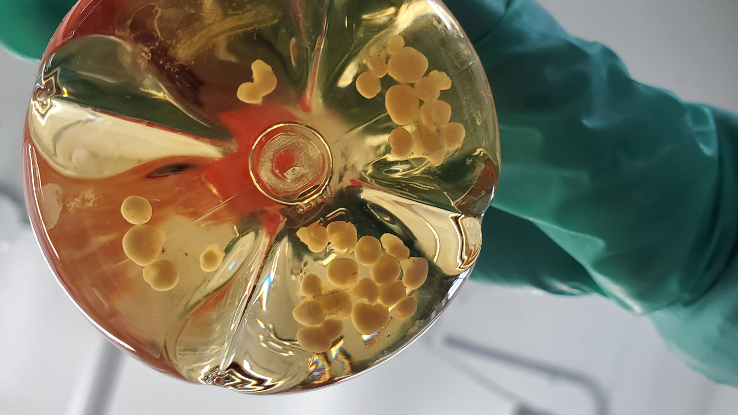 Figure: Fluorescence micrograph of transplanted human neurons (green, red) wiring into surrounding mouse brain tissue (blue). Source: Science News / S. Pasca lab, Stanford University
Building Neural Roads in a Living Brain: What Petri Dishes Cannot Do
To unravel how brain diseases take root, medicine took its first major step over a decade ago. In the early 2010s, an Austrian team showed that human stem cells could self-organize in a dish into miniature 3-D tissue clusters known as brain organoids. These pea-sized balls of cells generated rudimentary structures and brought renewed optimism to neuroscience.
Yet miniature brains in petri dishes quickly hit a biological wall. Without a functioning circulatory system to deliver nutrients and oxygen, and lacking electrical signals from other organs, human brain cells stall in their development once they reach a certain size.
To give these neurons a dynamic, living environment, Pașca’s team made a bold attempt in 2022. They transplanted human brain organoids into newborn rats. The human cells tapped into the rats’ blood vessels and survived. However, because rat neurons mature far faster than human neurons, the human cells could only occupy about one-third of one side of the rat’s cortex, capping their growth before extensive circuits could form.
Moving from test tubes to living animals required carving out dramatically more room inside the host’s skull.
Making Room in the Mouse Skull: Genetic Editing Clears the Cortex
For this latest study, the Stanford researchers adopted a far more radical strategy: genetically engineering a mouse model specifically designed to host human brain tissue.
These mice were bred without most of their cerebral cortex—the outer mantle responsible for higher-order cognition—and their hippocampus, the memory processing hub. Together, these missing structures account for roughly half of the animal’s total brain volume. Remarkably, despite missing half of their forebrain, the newborn mice maintained basic vital signs and proved “surprisingly functional.”
The resulting cavity served as a natural incubator for human tissue. Researchers transplanted brain organoids grown from healthy human donor stem cells into the void.
Out of 29 delicate microsurgeries, 25 transplants successfully took hold—an 86% success rate that underscores the stability and reproducibility of the surgical protocol.
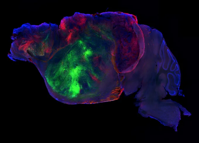 Figure: Micrograph showing neural connections from the same study. Source: Science News / S. Pasca lab, Stanford University
90% of the Cortex Replaced by Human Cells: Rare Neurons Flourish in Vivo
Once nestled inside the mice’s skulls, the human cells exhibited astonishing growth. Over the ensuing three months, the transplanted human tissue expanded nearly fivefold in volume.
Ultimately, human neurons came to dominate more than 90% of the mouse cortex. They hijacked the host’s blood vessels for oxygen and nutrients and wired extensively into the mouse’s existing nervous system, forging a true chimera—a functional hybrid of two distinct species.
Even more remarkably, the living environment coaxed the human cells into differentiating into specialized, rare neuron types that have proven virtually impossible to generate in culture dishes.
Although the transplanted tissue has not yet replicated the canonical six-layered architecture of the human cortex, it formed active, functional human neural circuits. Liberated from the confines of a petri dish, human cells became an integrated, signal-transmitting network inside a living mammal.
Modeling Cerebral Palsy: Patient-Derived Cells Bring Precision to Drug Testing
With the chimeric model established, the researchers immediately put it to work. They subjected the grafted mice to oxygen deprivation, successfully modeling the brain injury underlying cerebral palsy in premature infants.
Following hypoxia, the human brain tissue inside the mice displayed cellular hallmarks of injury identical to clinical cerebral palsy. At the same time, the mice developed motor deficits, exhibiting gait and limb coordination impairments that closely mirror human symptoms.
This breakthrough provides an unprecedented weapon against brain disorders. Promising drug candidates tested in conventional lab mice frequently fail in human clinical trials because rodent neurons react fundamentally differently from human neurons.
Now, scientists can harvest skin cells directly from patients suffering from frontotemporal dementia—a devastating disorder affecting personality and language—or genetic forms of autism. By reprogramming those cells into stem cells and growing disease-specific organoids to implant into mice, researchers finally have a platform to test targeted therapies against human neurons inside a living animal.
Will They Take Over the World? Bioethicists Draw the Behavioral Line
The revelation that human cells can usurp 90% of a mouse’s cortex naturally sparked public discussions about ethical boundaries. H. Isaac Chen, a neurosurgeon and organoid researcher at the University of Pennsylvania, noted that the extent of humanization reaches new heights.
To navigate these uncharted waters responsibly, Pașca’s team consulted an independent ethics committee chaired by bioethicist Insoo Hyun of the National University of Singapore. The panel established a clear benchmark: the secular ethical boundary depends not on the percentage of human cells present, but on whether those cells fundamentally alter the animal’s cognitive capabilities and behavior.
Rigorous cognitive and motor testing showed that the chimeric mice performed somewhere between normal mice and brain-injured mice lacking a cortex. They displayed no enhanced intelligence, no superhuman problem-solving skills, and no traces of higher consciousness.
As the research team noted, although most of their cortex is human, these rodents remain ordinary lab mice—far more Pinky and the Brain’s Pinky than Brain, with no designs on world domination.
It took over a decade of scientific perseverance to bridge the divide between cell cultures and living organisms. By clearing out half of a mouse’s native brain and allowing human neurons to colonize 90% of the cortex, neuroscience now possesses its first living window into human neural circuits, capable of recreating human disease from patient cells and testing lifesaving therapies.
Reference Links:
- Science News report
- Nature paper