0.25 Seconds Before Speaking, a Blueprint Forms Above the Left Ear
Walk into a corner coffee shop in the morning and casually ask the barista for a hot Americano. From the moment the thought forms to the instant your lips part and shape the words, the process feels as seamless and automatic as a reflex. Most people assume speaking is like hitting a play button—words tumbling out in direct sync with thought.
A neurosurgery team at the University of California, San Francisco (UCSF) has discovered that the reality is far more intricate. Roughly 0.25 seconds before you articulate the very first syllable, a small patch of cortex just above and behind your left ear begins firing. A quarter of a second is about half the duration of a blink, yet in that lightning-fast window, the brain has already mapped out a complete structural blueprint for the word.
Edward Chang, the lead researcher and chair of neurological surgery at UCSF, stated at the Society for Neuroscience meeting in November 2026: “Every single word that you’re going to say today started in this tiny region above your left ear, roughly a quarter-second before you said it.” Medically, this elusive area is the left posterior superior temporal gyrus (lpSTG)—a patch of cortex tucked behind and above the left ear, measuring somewhere between the size of a pea and a coin. Long categorized strictly as part of receptive auditory territory, it has now been revealed as a decisive starting point for vocal articulation.
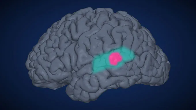 Figure: Brain region model showing Wernicke’s area in blue and lpSTG in pink. Source: Science News (Chang Lab)
Overturning a Century and a Half of Textbook Dogma
For nearly 150 years, medical curricula split the human language network into two neat compartments. In 1861, French physician Paul Broca identified Broca’s area, concluding that it exclusively orchestrated the motor mechanics of speaking with the tongue and vocal cords. In 1874, German neurologist Carl Wernicke identified Wernicke’s area on the left side of the brain, leading scholars to believe it was dedicated solely to receiving acoustic signals and comprehending language.
“Broca speaks, Wernicke listens”—this dichotomy became gospel in biology textbooks across generations. Clinicians and scientists envisioned language processing as a strict, one-way conveyor belt: sound waves entered through the ears and were decoded in Wernicke’s area, passing the baton over to Broca’s area only when it was time to command the vocal tract. That rigid division of labor guided neuroanatomy for a century and a half.
The paper published by Chang’s team in Science uproots this long-standing divide. lpSTG, nestled at the core of Wernicke’s area, fires robustly when listening to others, but it fires just as vigorously when preparing to speak oneself. As Leigh Hochberg, a brain-computer interface specialist at Brown University, put it: “Every brain area in the textbook that was labeled as doing just one thing, over the next decade, we’re going to learn that it does more.”
28 Surgical Patients and Intracranial Grids Pinpoint the Spark
Capturing neural flickers within a 250-millisecond window inside living human tissue presents severe experimental obstacles. Standard scalp EEG caps, muffled by the thickness of the scalp and skull, capture electrical signals as blurry as a landscape viewed through frosted glass. The researchers turned to 28 neurosurgical patients undergoing craniotomies for epilepsy or brain tumors.
To map critical speech centers and preserve language faculties during surgery, surgeons placed high-density electrocorticography (ECoG) electrode grids directly onto the surface of the cortex. In precious windows while patients were awake and cooperative during monitoring, researchers asked them to read various words displayed on a screen. Resting flush against the brain tissue, the electrodes recorded the minute electrical spikes of hundreds of neurons at millisecond resolution.
The data revealed a striking pattern. When participants saw short, single-syllable words like “cat,” lpSTG registered a modest ripple of electrical activity. But when they prepared to read a multi-syllable word like “caterpillar,” electrical discharges in the region surged several times over. The more structurally complex and practiced the word, the earlier and higher the amplitude of activation in this coin-sized patch of tissue before vocalization began.
This indicates that the region packages the structural blueprint of the entire word all at once, processing it as a unified semantic and phonetic unit. Across all 28 subjects, the intracranial recordings proved remarkably consistent, ruling out individual anomaly.
A Micro-Current Interruption Freezes Words on the Tongue
Recording neural firing alone does not prove causality; the region could theoretically be a bystander reflecting upstream preparation. Alexander Silva, a bioengineer at UCSF, designed an elegant electrical stimulation control experiment. Researchers delivered brief, tiny pulses of current to lpSTG at distinct millisecond intervals leading up to speech.
The outcome was definitive. When the micro-current was applied within the critical 0.25-second window prior to utterance, participants abruptly stalled mid-phrase, as if their speech apparatus had momentarily lost its roadmap. Yet if the very same electrical pulse was delivered immediately after the speaker uttered the first syllable, speech flowed uninterrupted.
This direct disruption offers conclusive proof: without the advance blueprint drawn by this area, downstream motor commands cannot proceed smoothly. Jeffrey Binder, a neurologist at the Medical College of Wisconsin, noted that this body of physiological and electrophysiological evidence is highly compelling, firmly placing this cortical patch within the command chain of speech generation.
An Upstream Decoding Target for Paralyzed Patients
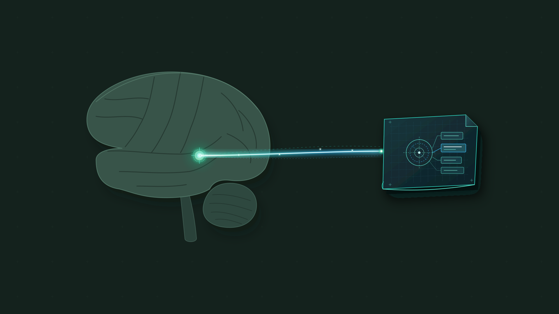
 Figure: Conceptual illustration depicting the neural pathways connecting speech comprehension and production. Source: National Geographic
This foundational scientific finding carries immediate implications for clinical engineering. Most current speech brain-computer interfaces (BCIs)—systems creating a direct communications link between neural tissue and external hardware—place sensor arrays over the motor cortex, the region responsible for sending physical motor commands to the larynx, tongue, and facial muscles. Engineers decode those muscle intentions and reconstruct what the patient intends to say.
Patients with severe stroke or amyotrophic lateral sclerosis (ALS), however, frequently suffer damage to these very motor execution pathways. When motor cortical circuits atrophy or scar, downstream sensors struggle to pick up distinct motor commands. Because lpSTG sits upstream of motor execution, it captures the abstract linguistic and word-level representations directly, bypassing the need for intact muscular control signals.
The research team has already tested novel implants in two volunteers living with paralysis and anarthria. Using signals recorded exclusively from lpSTG, the electrode arrays simultaneously decoded both the sentences participants heard and the words they intended to speak. Where neuroengineers previously had to implant multiple sensor arrays across separate functional zones, a single coin-sized cortical site can now bridge both sides of the linguistic loop.
This long-overlooked region behind the ear redraws our understanding of human speech architecture. By uniting receptive listening and active articulation within a shared neural circuit, it offers brain-computer interfaces a direct gateway into the inner speech of those who cannot talk. As intended words are registered at their cognitive inception, the fundamental beginnings of human speech come into sharper focus.
References:
- Science News coverage
- National Geographic feature analysis
- Original paper in Science