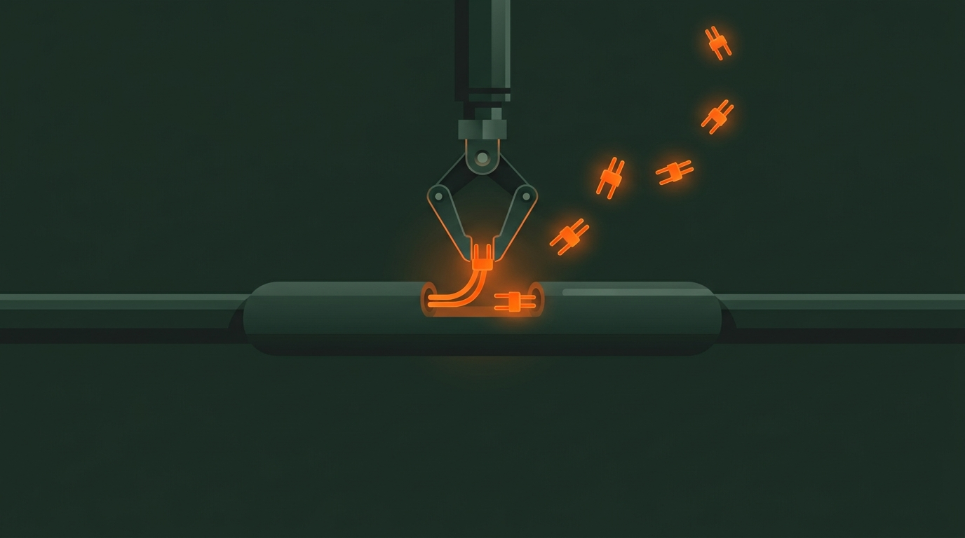
A Blackout in the Deep: When Retinal Nerves Lose Their Power
In everyday life, a tripped circuit breaker instantly paralyzes home appliances, plunging rooms into darkness. For individuals who lose their vision to trauma or cerebral hemorrhage, a similarly desperate blackout occurs inside the eye.
Clear vision relies on photoreceptor cells in the retina—the light-sensitive membrane lining the back of the eyeball—and the optic nerve, the biological cable connecting the eye to the brain to transmit visual signals. When severe trauma interrupts local blood flow, oxygen deprivation starves these nerve cells of energy, causing them to cease functioning.
In a study posted on a preprint server on August 10, 2026, researchers at the Icahn School of Medicine at Mount Sinai in New York executed a clinical procedure never before attempted in the human eye. A female patient, who had been blind for two months following a brain hemorrhage, received a specialized treatment designed to deliver energy directly into her eyes.
From Thigh to Eyeball: Delivering Backup Batteries to Damaged Cells
Nerve cells require immense amounts of energy due to their dense populations of mitochondria—the microscopic cellular power plants that produce energy. Mitochondria are tiny, typically measuring just 0.5 to 1 micrometer in diameter, roughly one-hundredth the width of a human hair. A healthy neuron often clusters hundreds or thousands of these micro-generators, working around the clock to synthesize chemical energy.
When the optic nerve is damaged, internal mitochondria are the first to degrade, causing the entire nerve cell to fall dormant from energy exhaustion. Rather than attempting high-risk stem cell transplants, the research team opted to borrow healthy generators from the patient’s own body. They extracted active mitochondria from her thigh muscle—a high-intensity tissue with abundant, exceptionally healthy mitochondrial reserves.
During the procedure, surgeons used an ultra-fine needle to slowly inject these autologous mitochondria into the vitreous fluid—the transparent gel filling the eyeball that maintains its shape—in both eyes. Once injected, the floating micro-generators crossed cellular boundaries and were taken up by retinal cells, resupplying critical energy to dying neurons.
 Figure: False-color electron micrograph of a mitochondrion, highlighting its outer membrane and inner cristae structure. Source: Nature / K. R. Porter/SPL
Four Weeks of Light Sensing: Physiological Miracles and Therapeutic Limits
Prior to surgery, the patient had been completely blind for two months. Her pupils showed no constriction response to bright light, and ocular physiological signals were entirely silent. Following the intravitreal mitochondrial injection, however, her dormant eyes demonstrated a clear physiological response.
Post-operative testing revealed that her unreactive pupils regained an active constriction response to light—a reaction that persisted for a full four weeks. In a severely damaged eye, four weeks of pupillary light response indicated that the injected mitochondria successfully reactivated dormant neural circuits.
Low-vision evaluations showed that the patient’s left eye began perceiving general shapes and shadows, though the trial did not restore functional vision for daily life. After four weeks, the pupillary response gradually diminished. The project lead explicitly noted that while the team cannot claim clinical efficacy, they observed remarkable ocular physiological activation at the physical level.
 Figure: Cross-sectional view of mitochondrial ultrastructure, illustrating the core machinery for cellular energy delivery. Source: Nature
Awakening Dormant Cells: The First Step in Safety Verification
Previous mouse studies demonstrated that injecting mitochondria into the vitreous cavity protects the optic nerve, significantly increasing neuron survival after injury. In clinical medicine, mitochondrial transplantation has seen preliminary exploration in cardiac and neurological surgeries, but injection into a highly sensitive, enclosed organ like the eye marks a global first.
Mitochondrial experts at the Institute of Molecular and Clinical Ophthalmology Basel (IOB) noted that the primary value of this trial lies in proving the relative safety of intravitreal autologous mitochondrial injections. Over the observation period, the patient experienced no severe immune rejection or inflammatory reactions, laying a crucial safety foundation for future research.
While a single-case study provides limited statistical weight, it opens a novel perspective for treating neurodegenerative eye diseases. Confirming that exogenous mitochondria can cross cell membranes and be absorbed by neurons suggests that cell-energy supplementation could buy a critical therapeutic window for optic nerve preservation.
Replacing Organs vs. Repairing Power Plants: A New Era in Blindness Therapy
Injecting a patient’s own mitochondria into the eye shifts the paradigm of blindness treatment from tissue replacement to repairing cellular power plants. Traditional treatments for severe ocular conditions often aim to replace damaged cells entirely or implant artificial lenses—approaches plagued by high engineering complexity and a risk of disrupting intricate neural networks.
Awakening damaged retinal neurons with healthy mitochondria provides a gentler, cell-level therapeutic path. Even if full clarity of vision remains out of reach for now, transferring energy from thigh muscle to eyeball highlights compelling new possibilities in cellular repair.
Reference Links:
- Nature News Report
- Icahn School of Medicine at Mount Sinai Preprint Study