
When You Cough, Billions of Airway Brushes Are Working Overtime
When you catch a cold and start coughing, few people wonder what is actually happening deep inside the trachea. Lining the inner walls of our windpipe and bronchi is a round-the-clock biological clearance system. The surface of the human respiratory tract is blanketed by billions of microscopic, hair-like projections known as cilia.
Each cilium measures only about 200 nanometers in diameter—less than one-five-hundredth the thickness of a human hair. In a healthy individual, these cilia beat ten or more times per second in coordinated, wave-like rhythms. Functioning as miniature biological brooms, they continuously propel mucus, dust, and pathogens toward the throat, where they are eventually expelled by coughing or swallowed.
When this synchronized rhythm breaks down, the airway is in serious jeopardy. Primary ciliary dyskinesia (PCD)—a rare genetic respiratory disorder where the cellular brushes fail to beat normally—is a prime example. In PCD patients, the frequency and coordination of ciliary motion falter. Mucus and trapped foreign particles cannot be cleared, leaving affected children suffering from persistent, recurrent respiratory infections from infancy onward.
Invisible in Still Stills: A 10-Second Clip Pinpoints the Breakdown
On September 15, 2026, the winners of the 16th Nikon Small World in Motion competition were officially unveiled. Dr. Ning Xu, an optical engineering researcher from the Department of Precision Instrument at Tsinghua University, claimed first place. His winning submission documented the live movement of cilia in a pediatric PCD patient, standing out among 346 entries from 40 countries.
The winning footage is far from traditionally picturesque. The cilia on the patient’s respiratory epithelial cells twitch erratically, shuddering and pausing in localized clusters. Yet this abnormal kinetic pattern is the exact pathological source of the child’s chronic infections.
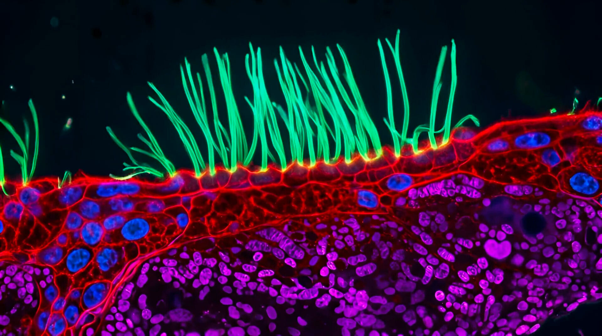 Image: Abnormal beating of airway cilia in a child with PCD. Source: Nikon Small World / Dr. Ning Xu, Tsinghua University
Historically, clinical pathology has relied heavily on static tissue biopsies. Under a conventional still photograph, the physical structure of a patient’s cilia often appears completely intact, making a definitive diagnosis exceptionally challenging for clinicians relying on slice photography alone. Dr. Xu noted following the announcement that understanding this condition requires observing dynamic motion continuously over roughly 10 seconds—still images simply do not tell the whole story.
No Fluorescent Dyes: Illuminating Living Cells with Structured Light
Recording the high-speed trajectories of living cilia under a microscope is a formidable engineering feat. While electron microscopes offer immense magnification, they require dehydrated, fixed samples, rendering them incapable of imaging living cells. Standard fluorescence microscopy requires injecting chemical stains and illuminating them with high-intensity light, which risks phototoxic damage to fragile living cells and can disrupt the natural beating cadence of cilia.
Dr. Xu’s team turned to diffractive super-resolution microscopy—an imaging approach that surpasses the classical diffraction limit of light by precisely modulating illumination, allowing crisp visualization of living cellular structures without staining. Because no chemical labels are required, researchers can continuously record living specimens in their natural, physiological state.
On the hardware side, the system integrates a Digital Micromirror Device (DMD)—a micro-electro-mechanical chip composed of an array of microscopic mirrors that can steer light beams with microsecond precision. By projecting tailored light patterns onto the sample, the DMD minimizes total photon exposure while maximizing contrast. Paired with a 100x objective lens, the setup resolves the rapid, subtle fluttering of living cilia, turning ephemeral cellular dynamics into unequivocal diagnostic evidence.
Wonders of the Microverse: Highlights from the Competition
This year’s competition drew 346 video submissions from around the globe, with researchers utilizing cutting-edge optics to reveal fleeting biological events that usually escape human sight.
Nguyen Nam Nhat from Vietnam took second place with a mesmerizing video capturing a tiny nematode worm and a single-celled Dileptus protist exploring and hunting within a microscopic water droplet.
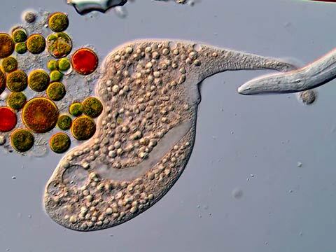 Image: A nematode and single-celled Dileptus navigating the microverse. Source: Nikon Small World / Nguyen Nam Nhat
Third place went to Benedikt Pleyer of Germany, who filmed the delicate swimming motion of a jellyfish larva suspended in a drop of water. Fourth place was awarded to Andrew Moore of the Howard Hughes Medical Institute (HHMI) in the United States for capturing synchronous cell division in adjacent cells. Fifth place went to Patrick Hickey from the United Kingdom, who recorded the streaming motion of mitochondria and chloroplasts inside plant leaf cells.
As Dr. Xu reflected, seeing cilia in motion is deeply intuitive—it allows anyone to instantly realize that vital biological processes are occurring within our bodies. Micro-cinematography is not merely a specialized scientific tool; it has evolved into a universal visual language that connects the public with microscopic biology.
Motion as Evidence: How a Microscopic Lens Reimagines Diagnosis
This 10-second recording is far more than a triumph of optical engineering. It exemplifies a long-overlooked diagnostic philosophy: motion itself is evidence. In traditional medical diagnostics, clinicians are conditioned to search for morphological defects—masses, structural tears, or genetic mutations.
Yet within the human body, countless vital physiological functions depend on high-frequency, precision mechanical movement. When the kinetic rhythm falls out of sync, structurally intact cells still fail to do their jobs. And the pathology wrought by motion disorders leaves few traces in static snapshots.
Some diseases cannot be captured by photographs; they require video. Dynamic videography introduces the dimension of time into cellular examination, transforming invisible cellular rhythms into incontrovertible visual evidence. For disorders like PCD—diseases defined by motion—these 10 seconds of motion are the diagnosis.
References:
- 16th Annual Nikon Small World in Motion Competition Winners
- Ars Technica Coverage of Nikon Small World in Motion