Why 99% of Psychiatric Drugs Fail in Animal Trials: Mice Simply Don’t Get Depressed
Many patients living with depression or anxiety know the ordeal intimately. After receiving a prescription, they often spend weeks or even months testing different medications just to find an effective combination. Psychiatric drugs have long suffered from among the highest failure rates in clinical trials; vast numbers of candidate compounds that demonstrate remarkable promise in laboratory benchmarks ultimately fail when tested in human patients.
This persistent bottleneck stems from a formidable biological barrier: the brain architecture of experimental animals is fundamentally distinct from our own. Standard laboratory mice cannot develop psychiatric disorders unique to human beings. Complex conditions such as depression and autism involve the pathological divergence of higher-order cortical circuits. When scientists evaluate prospective compounds in mice, they merely capture the responses of a rodent nervous system—data incapable of illuminating the true biology of the human brain.
To obtain more biologically relevant observations, scientists have historically relied on culturing cell clusters in petri dishes. Yet cerebral organoids floating in nutrient broth lack a vascular blood supply and cannot replicate the complex electrical dynamics of a functioning, living brain. Identifying a physiological host capable of sustaining authentic human neural circuits in vivo has long been the central challenge of modern brain disease research.
Genetic Surgery Clears Space for Transplanted Human Skin Cells to Rebuild the Cortex
A research team at Stanford University has now published a breakthrough study in the journal Nature. The researchers began by genetically engineering mice to induce an apallial state during embryonic development—a physiological condition where targeted genetic disruption prevents the cerebral neocortex and hippocampus from developing. This genetic surgery created a substantial growth void inside the mouse skull where the cortex would normally reside.
Next, the team reprogrammed human skin cells into induced pluripotent stem cells and differentiated them into cortical organoids—miniature human brain tissues grown in vitro. During early postnatal stages, researchers transplanted these human neural clusters directly into the cranial void of the mice. These animals are collectively designated “xenocortical mice”—experimental mice whose cerebral cortices are predominantly populated and functionally replenished by human neurons.
As the mice matured, the transplanted human cells underwent spontaneous, self-guided development within the living host. Human neurons not only filled the void completely, but also organized into layer 5 pyramidal neurons—the principal projection neurons in the cerebral cortex responsible for relaying long-range signals. Remarkably, specialized cell types previously observed exclusively in the human brain also spontaneously emerged and formed within the rodent skull.
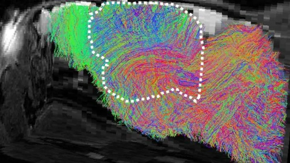 Figure: Brain scan of a mouse with transplanted human brain cells; colored lines indicate neural pathways. Source: Stanford
Normal Electrical Firing in Vivo: Six-Month-Old Mice Retain Normal Motor Skills
Crucially, these cross-species grafts did not remain isolated, dormant islands of tissue. Using calcium imaging—a fluorescent recording technique that tracks real-time neuronal firing in living tissue—the team discovered that the transplanted human neurons formed functional synaptic connections with neighboring mouse brain regions. These human neurons successfully received sensory signals from the mouse and generated coordinated, rhythmic electrical firing patterns.
The typical lifespan of a mouse is only about two years. The researchers tracked the transplanted mice over a six-month window, effectively monitoring their developmental trajectory across a full quarter of the animal’s lifetime. Throughout this extended duration, the human neurons maintained their characteristically protracted maturation timeline, developing at a natural human pace without succumbing to distortions from the mouse’s accelerated metabolism.
In behavioral assessments, the xenocortical mice demonstrated robust functional adaptation. Their baseline motor capabilities remained virtually identical to those of control littermates, displaying neither artificial intelligence enhancements nor abnormal behavioral traits. Researchers detected only subtle differences in fine motor limb coordination, offering direct physical evidence that the human neural circuits were actively participating in modulating the animal’s movement.
Human Circuits in Lab Mice: Scientists Confront the Ethical Frontier
The fact that human brain cells can take over and operate the cortex inside an animal skull has triggered deep reflection across the bioethics community. Dr. Sarah Chan, a bioethicist at the University of Edinburgh, pointed out that while the mice currently show no indications of human-like cognition or reflective thought, the research compels humanity to re-evaluate the technical and ethical thresholds surrounding animal cognitive alteration. Assessing the conscious and perceptual experiences of such chimeric models represents an urgent frontier for biomedical ethics.
Dr. Ilary Allodi, a neuroscientist at the University of St Andrews, noted that the mouse brain effectively acted as a “natural incubator” for the graft. Although the transplanted cortex exhibited a somewhat disordered anatomical architecture, its capacity for self-assembly vastly exceeded expectations. Other researchers have cautioned that strict ethical oversight and procedural complexity may restrict how rapidly this technology can be scaled.
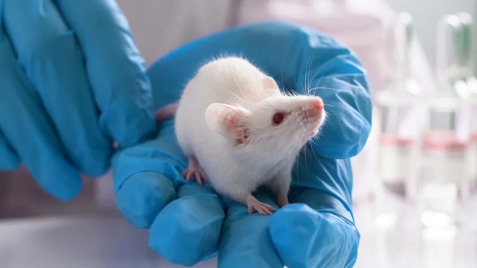 Figure: White laboratory mouse resting on a hand wearing a blue protective glove. Source: BBC
The Stanford team underscored that the explicit purpose of this technology is to construct precise disease models while keeping animal cognition stable and predictable. Conducting these experiments under stringent, transparent oversight is essential to ensuring that these discoveries can be translated safely into clinical therapies.
Living Circuits Dismantle Drug Discovery Barriers: A Realistic Human Neural Model Arrives
The breakthrough from Stanford provides a transformative platform for drug screening in disorders such as autism, epilepsy, and developmental neuropathologies. In the past, pharmaceutical teams had no choice but to test candidate molecules either in isolated cell cultures or in purely rodent models. Today, they can directly monitor how prospective therapeutic compounds regulate authentic human neural circuits operating within a living physiological host. Such in vivo testing promises to slash the substantial trial-and-error costs incurred during early clinical phases.
For psychiatric medicine—a field long trapped in a research-and-development winter—this technology fills a profound methodological void. Evaluating candidate molecules against active human neural circuits in vivo will enable researchers to identify ineffective or toxic compounds far earlier, potentially rescuing psychiatric medicine from its notoriously high clinical failure rates.
By allowing human neurons to grow into a functional cortex within living mice, brain disease research can for the first time evaluate authentic human neural circuits in a living system. The fundamental reason 99% of psychiatric drugs have failed in animal trials is that mice do not suffer from human mental illnesses. This mouse, endowed with functional human cortical circuits, has opened the decisive breakthrough to finally solve that problem.
Reference Links:
- BBC News Report
- Nature Paper